You have no items in your shopping cart.
KO/KD
Validated
Validated
Description
Research Area
Cell Biology
Images & Validation
−Item 1 of 5
| Tested Applications | ELISA, IHC, IP, KO/KD Validated, WB |
|---|---|
| Dilution Range | ELISA: 1:4,000 - 1:20,000, IHC: 1:1,000, IP: 10-30ul, WB: 1:500 – 1:3000 |
| Reactivity | Mouse |
| Application Notes |
Key Properties
−| Antibody Type | Primary Antibody |
|---|---|
| Host | Rabbit |
| Clonality | Polyclonal |
| Isotype | Antiserum |
| Immunogen | This whole rabbit serum was prepared by repeated immunizations with a recombinant fusion protein from amino acids 601-1115 of mouse deltaN PERK. |
| Target | Eif2ak3 |
| Purity | This antiserum is directed against PERK and reacts with the PERK from mouse tissues. Reactivity to other species is unknown. |
| Conjugation | Unconjugated |
Storage & Handling
−| Storage | Store vial at -20° C prior to opening. Aliquot contents and freeze at -20° C or below for extended storage. Avoid cycles of freezing and thawing. Centrifuge product if not completely clear after standing at room temperature. This product is stable for several weeks at 4° C as an undiluted liquid. Dilute only prior to immediate use. |
|---|---|
| Form/Appearance | Liquid (sterile filtered) |
| Buffer/Preservatives | 0.01% (w/v) Sodium Azide |
| Concentration | 80 mg/mL |
| Expiration Date | 12 months from date of receipt. |
| Dry Ice Shipping | Please note: This product requires shipment on dry ice. A dry ice surcharge will apply. |
| Disclaimer | For research use only |
Alternative Names
−rabbit anti-PKR-like Endoplasmic Reticulum Kinase Antibody, rabbit anti-PERK Antibody, PKR-like endoplasmic reticulum kinase, PERK, Eukaryotic translation initiation factor 2-alpha kinase 3
Similar Products
−- Item 1 of 10
Phospho-PERK (Thr982) Rabbit Polyclonal Antibody [orb500026]
FC, ICC, IF, IHC-Fr, IHC-P
Rabbit
Human, Mouse, Rat
Rabbit
Polyclonal
Unconjugated
100 μl, 50 μl, 200 μl - Item 1 of 8
Phospho-PERK (Thr980) Rabbit Polyclonal Antibody [orb506147]
FC, IF, IHC-Fr, IHC-P, WB
Bovine, Canine, Porcine, Rabbit
Human, Mouse, Rat
Rabbit
Polyclonal
Unconjugated
50 μl, 100 μl, 200 μl - Item 1 of 7
PERK Rabbit Polyclonal Antibody [orb13645]
FC, IF, IHC-Fr, IHC-P, WB
Mouse, Rat
Human, Mouse, Rat
Rabbit
Polyclonal
Unconjugated
50 μl, 100 μl, 200 μl - Item 1 of 4
PERK rabbit pAb Antibody [orb770922]
ELISA, IF, IHC, WB
Human, Mouse, Rat
Polyclonal
Unconjugated
100 μl - Item 1 of 1
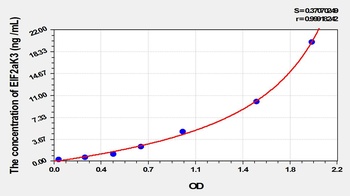
Mouse Eukaryotic Translation Initiation Factor 2 Alpha Kinase 3 (EIF2aK3) ELISA Kit [orb780866]
Mouse
0.32-20 ng/mL
0.11 ng/mL
96 T, 48 T


Quality Guarantee
Explore bioreagents carefree to elevate your research. All our products are rigorously tested for performance. If a product does not perform as described on its datasheet, our scientific support team will provide expert troubleshooting, a prompt replacement, or a refund. For full details, please see our Terms & Conditions and Buying Guide. Contact us at support@biorbyt.com.
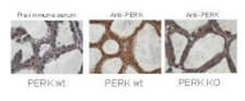
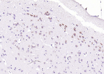
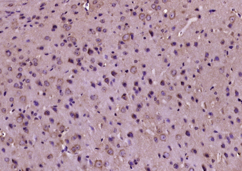
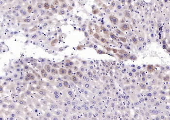
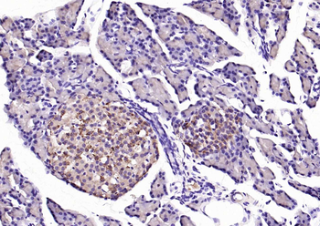
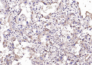
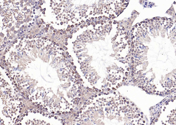
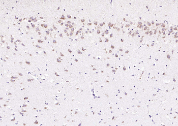
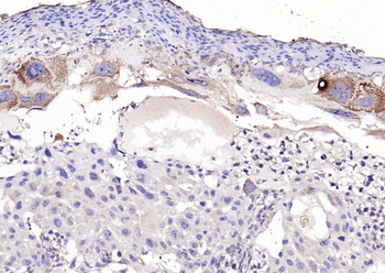
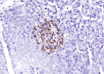
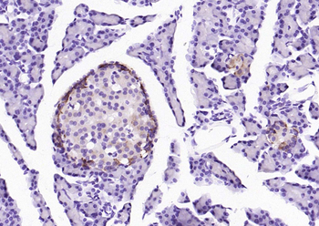
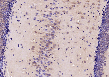
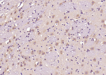
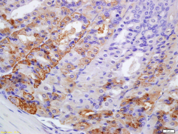
Immunohistochemistry staining of mouse mammary gland samples from lactating mice (L10) with Biorbyt's anti-PERK. Positive staining signal observed in wild type mouse sample with anti-PERK staining only (middle image), but not in the knock out mouse sample (right image) and pre-immune serum staining (left image) The anti-PERK was diluted 1:1000 in 5% goat serum in PBS and allowed to incubate for 2h at room temperature in a humidified chamber.
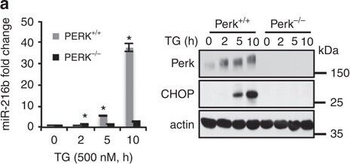
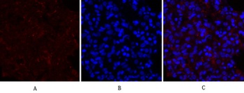
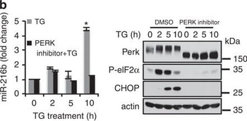
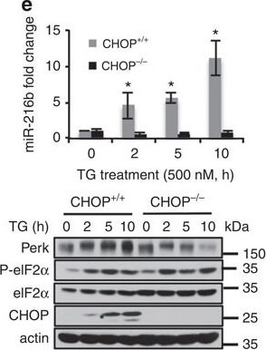
PERK-dependent miR-216b induction.(a) PERK+/+ and PERK−/− MEFs were treated with 500 nM TG for indicated times. MiR-216b was assessed by qPCR (left graph); PERK and CHOP were assessed by immunoblot (right). (b) MiR-216b levels were quantified by qPCR following exposure of cells to thapsigargin and a small-molecule PERK inhibitor (left). PERK, eIF2α-p and CHOP induction were assessed by immunoblot (right). (c–e) MEFs of the indicated genotype were treated with TG (500 nM) for indicated intervals. Protein extracts from these cells were immunoblotted for the proteins as indicated (lower panels) and miR-216b levels were quantified by qPCR (upper panels; n = 3). (f) CHOP−/− MEFs were transfected with vector or CHOP and 2 days later treated with TG (500 nM) for indicated intervals. Protein extracts from these cells were immunoblotted for CHOP and miR-216b levels quantified by qPCR (n = 3). (g) MiR-216b expression and (h) c-Jun mRNA levels were analysed in MMTV-Neu tumours from either PERK+/+ or a PERK−/− background. Data represent mean±s.d. of three independent observations. Statistical significance was analysed analysed by Student's t-test. (*P < 0.05, WT versus −/−).
PERK-dependent miR-216b induction.(a) PERK+/+ and PERK−/− MEFs were treated with 500 nM TG for indicated times. MiR-216b was assessed by qPCR (left graph); PERK and CHOP were assessed by immunoblot (right). (b) MiR-216b levels were quantified by qPCR following exposure of cells to thapsigargin and a small-molecule PERK inhibitor (left). PERK, eIF2α-p and CHOP induction were assessed by immunoblot (right). (c–e) MEFs of the indicated genotype were treated with TG (500 nM) for indicated intervals. Protein extracts from these cells were immunoblotted for the proteins as indicated (lower panels) and miR-216b levels were quantified by qPCR (upper panels; n = 3). (f) CHOP−/− MEFs were transfected with vector or CHOP and 2 days later treated with TG (500 nM) for indicated intervals. Protein extracts from these cells were immunoblotted for CHOP and miR-216b levels quantified by qPCR (n = 3). (g) MiR-216b expression and (h) c-Jun mRNA levels were analysed in MMTV-Neu tumours from either PERK+/+ or a PERK−/− background. Data represent mean±s.d. of three independent observations. Statistical significance was analysed analysed by Student's t-test. (*P < 0.05, WT versus −/−).
PERK-dependent miR-216b induction.(a) PERK+/+ and PERK−/− MEFs were treated with 500 nM TG for indicated times. MiR-216b was assessed by qPCR (left graph); PERK and CHOP were assessed by immunoblot (right). (b) MiR-216b levels were quantified by qPCR following exposure of cells to thapsigargin and a small-molecule PERK inhibitor (left). PERK, eIF2α-p and CHOP induction were assessed by immunoblot (right). (c–e) MEFs of the indicated genotype were treated with TG (500 nM) for indicated intervals. Protein extracts from these cells were immunoblotted for the proteins as indicated (lower panels) and miR-216b levels were quantified by qPCR (upper panels; n = 3). (f) CHOP−/− MEFs were transfected with vector or CHOP and 2 days later treated with TG (500 nM) for indicated intervals. Protein extracts from these cells were immunoblotted for CHOP and miR-216b levels quantified by qPCR (n = 3). (g) MiR-216b expression and (h) c-Jun mRNA levels were analysed in MMTV-Neu tumours from either PERK+/+ or a PERK−/− background. Data represent mean±s.d. of three independent observations. Statistical significance was analysed analysed by Student's t-test. (*P < 0.05, WT versus −/−).
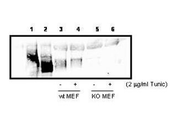
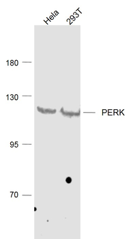
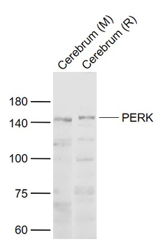
Western blot analysis using Biorbyt's anti-PERK to detect PERK in cell lysates. 300 µg PERK over-expressing 293T cell lysate (lanes 1 & 2), or 800 ug wild type (Lanes 3 & 4), and PERK knock out (lanes 5 & 6) MEF cell lysate were immunoprecipated with 15 µl anti-PERK, followed by western blotting with 1:1000 dilution of anti-PERK in 5% milk/TBST buffer. Lane 1, 293T cells over-expressing Myc-PERK wt, Lane 2, 293T cells over-expressing Myc-PERK K618A.
Documents Download
Datasheet
Product Information
Request a Document
Protocol Information
WB
Western Blot (IB, immunoblot)
IHC
Immunohistochemistry
ELISA
Enzyme-linked Immunosorbent Assay (EIA)
IP
Immunoprecipitation
Eif2ak3 Antibody (orb750495)

- 0.0
Based on 0 reviews
Participating in our Biorbyt product reviews program enables you to support fellow scientists by sharing your firsthand experience with our products.
Login to Submit a ReviewAvailable Sizes
Select a size below
Choose Conjugation or Carrier Free Version
Free Secondary Antibody (20 ul)0/0
Please add an antibody product to your cart first.