You have no items in your shopping cart.
Description
Research Area
Musculoskeletal & Connective Tissue Research
Images & Validation
−Item 1 of 10
| Tested Applications | ICC, IHC, WB |
|---|---|
| Reactivity | Human, Mouse |
Key Properties
−| Antibody Type | Primary Antibody |
|---|---|
| Host | Mouse |
| Clonality | Monoclonal |
| Isotype | IgG1 |
| Clone No. | RV204 |
| Conjugation | Unconjugated |
Storage & Handling
−| Storage | Maintain refrigerated at 2-8°C for up to 2 weeks. For long term storage store at -20°C in small aliquots to prevent freeze-thaw cycles. |
|---|---|
| Expiration Date | 12 months from date of receipt. |
| Disclaimer | For research use only |
Similar Products
−- Item 1 of 3
Vimentin Mouse Monoclonal Antibody [orb1474277]
IF, IHC, IP, WB
Human
Mouse
Monoclonal
Unconjugated
200 μl, 100 μl, 30 μl, 50 μl - Item 1 of 3
Vimentin Mouse Monoclonal Antibody [orb323201]
IF, IHC, WB
Human, Mouse, Rat
Mouse
Monoclonal
Unconjugated
30 μl, 100 μl, 200 μl, 50 μl - Item 1 of 2
- Item 1 of 3
Recombinant VIM Antibody / Vimentin [orb1151550]
IHC-P, WB
Human
Mouse
Recombinant
Unconjugated
100 μg, 20 μg - Item 1 of 1
VIM/Vimentin Rabbit Polyclonal Antibody [orb2955593]
ELISA, IHC, WB
Bacteria, Bovine, Canine, Gallus, Golden Hamster, Hamster, Human, Monkey, Mouse, Porcine, Primate, Rat, Sheep
Rabbit
Polyclonal
Unconjugated
50 μg, 100 μg


Quality Guarantee
Explore bioreagents carefree to elevate your research. All our products are rigorously tested for performance. If a product does not perform as described on its datasheet, our scientific support team will provide expert troubleshooting, a prompt replacement, or a refund. For full details, please see our Terms & Conditions and Buying Guide. Contact us at support@biorbyt.com.
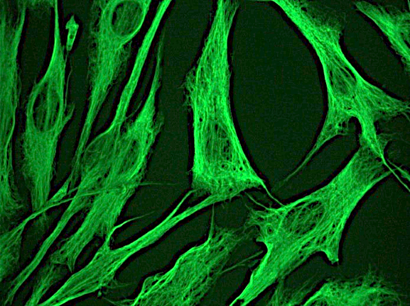
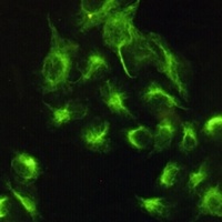
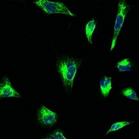
Indirect immunofluorescence staining of normal human dermal fibroblasts in tissue culture with orb669776 (diluted 1:250), showing the specific cytoskeletal pattern of vimentin intermediate filaments.
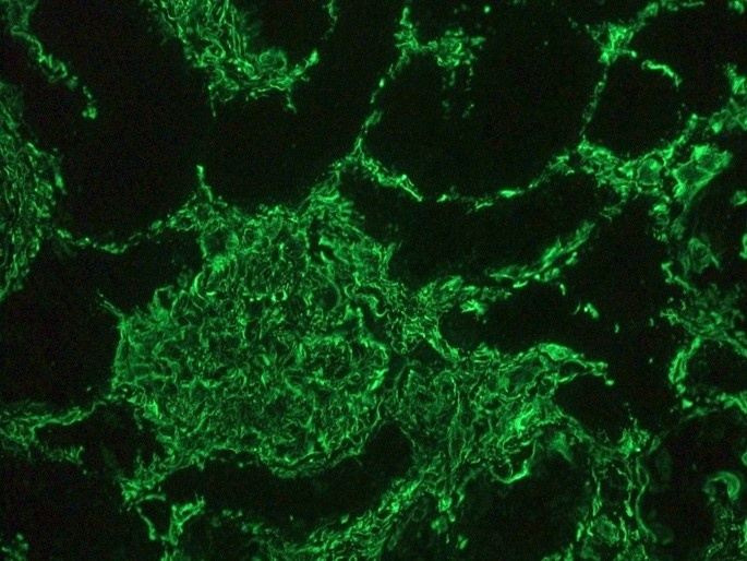
Indirect immunofluorescence staining of human kidney tissue section with orb669776 (diluted 1:1000), showing the specific pattern of vimentin in the mesenchymal cell types, such as fibroblasts in the connective tissue, podocytes, and endothelial cells in blood vessels. As expected, no reactivity is seen in the epithelial cell compartment
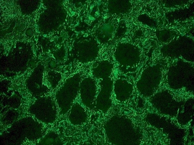
Indirect immunofluorescence staining of human kidney tissue section with orb669776 (diluted 1:1000), showing the specific pattern of vimentin in the mesenchymal cell types, such as fibroblasts in the connective tissue, podocytes, and endothelial cells in blood vessels. As expected, no reactivity is seen in the epithelial cell compartment
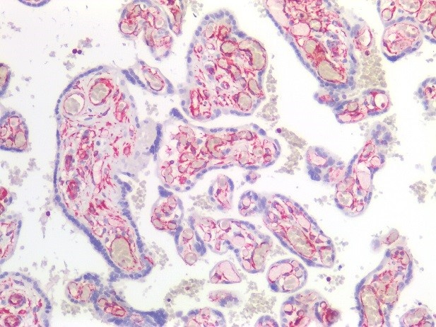
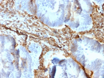
Immunostaining of human paraffin embedded tissue section of placenta with orb669776 (diluted 1:100), showing the specific pattern of vimentin in the mesenchymal cell types, such as fibroblasts in the connective tissue, and endothelial cells in blood vessels. As expected, no reactivity is seen in the epithelial cell compartment
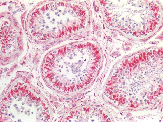
Immunostaining of human paraffin embedded tissue section of testis with orb669776 (diluted 1:100), showing the specific pattern of vimentin in the mesenchymal cell types, such as fibroblasts in the connective tissue, endothelial cells in blood vessels and Sertoli cells.
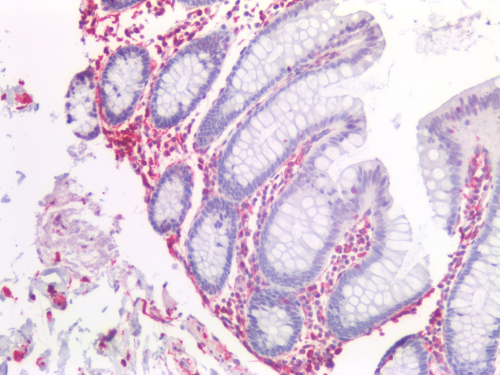
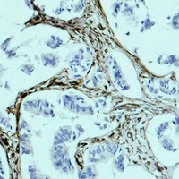
Immunostaining of human paraffin embedded tissue sections of human colon with orb669776 (diluted 1:100), showing the specific pattern of vimentin in the mesenchymal cell types, such as fibroblasts in the connective tissue, and endothelial cells in blood vessels. As expected, no reactivity is seen in the epithelial cell compartment.
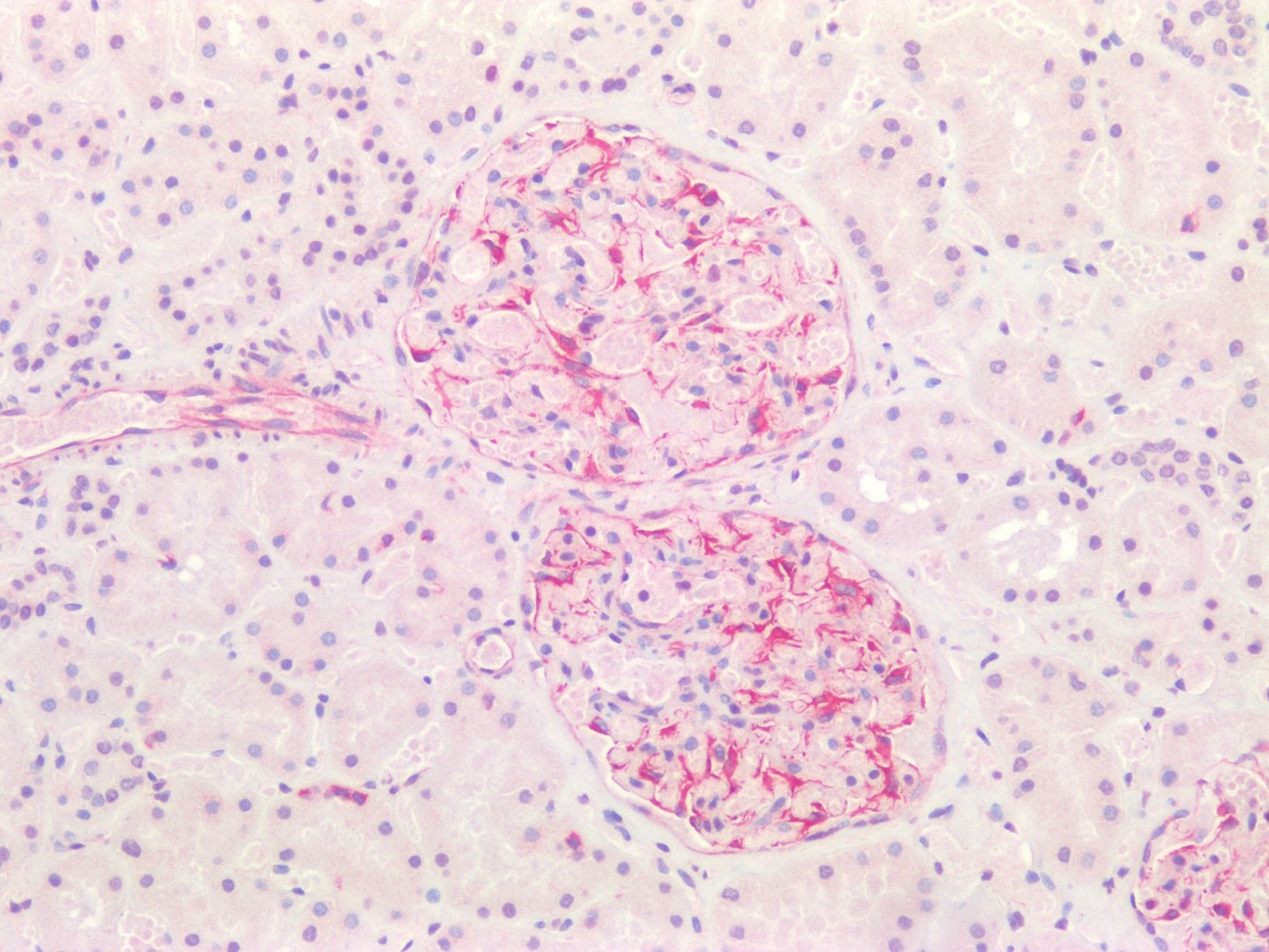
Immunostaining of human paraffin embedded tissue sections of human kidney with orb669776 (diluted 1:100), showing the specific pattern of vimentin in the mesenchymal cell types, such as fibroblasts in the connective tissue, and podocytes. As expected, no reactivity is seen in the epithelial cell compartment.
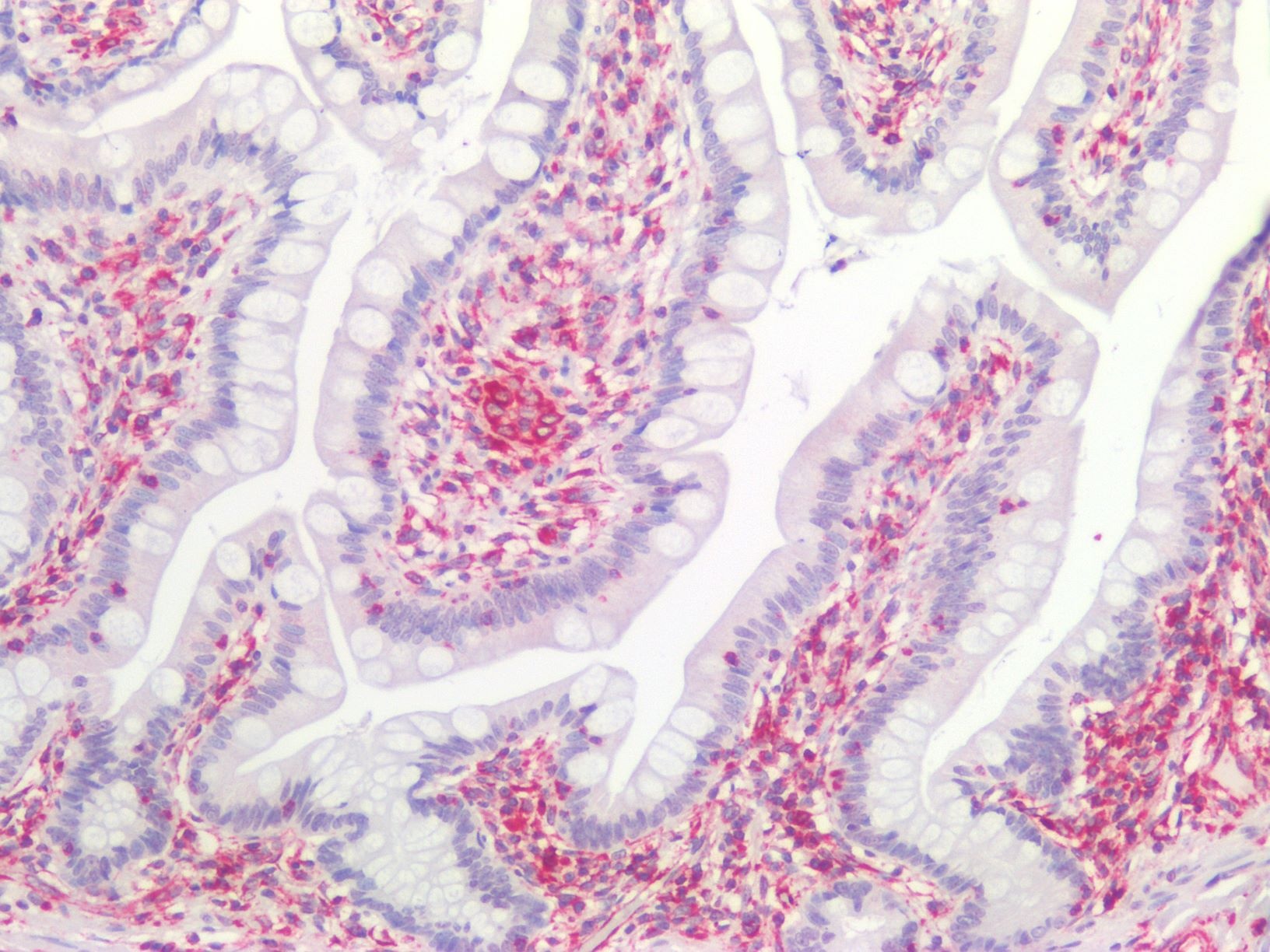
Immunostaining of human paraffin embedded tissue sections of human small intestine with orb669776 (diluted 1:100), showing the specific pattern of vimentin in the mesenchymal cell types, such as fibroblasts in the connective tissue. As expected, no reactivity is seen in the epithelial cell compartment.
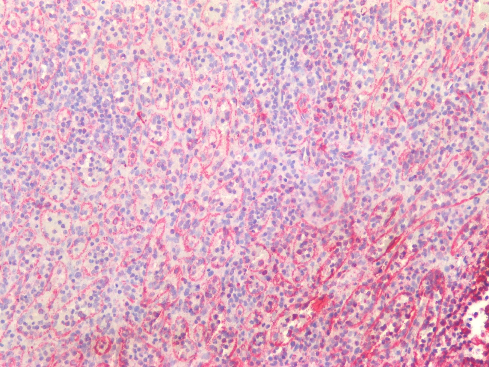
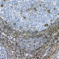
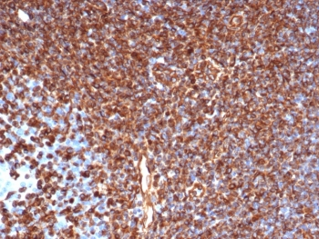
Immunostaining of human paraffin embedded tissue sections of human spleen with orb669776 (diluted 1:100), showing the specific pattern of vimentin in the mesenchymal cell types.
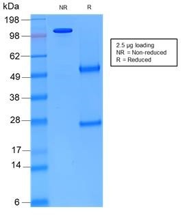
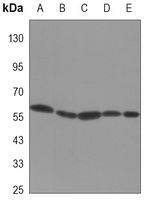
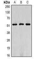
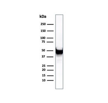
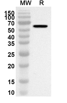
Non-reduced and reduced SDS-PAGE of orb669776 showing its purity.
Quick Database Links
UniProt
UniProt Details
− No UniProt data available
Documents Download
Datasheet
Product Information
Request a Document
Protocol Information
WB
Western Blot (IB, immunoblot)
IHC
Immunohistochemistry
ICC
Immunocytochemistry
Recombinant mouse vimentin Antibody (orb669776)

- 0.0
Based on 0 reviews
Participating in our Biorbyt product reviews program enables you to support fellow scientists by sharing your firsthand experience with our products.
Login to Submit a ReviewAvailable Sizes
Select a size below
Free Secondary Antibody (20 ul)0/0
Please add an antibody product to your cart first.