You have no items in your shopping cart.
Description
Research Area
Cell Biology
Images & Validation
−Item 1 of 4
| Tested Applications | IF, IHC-P, WB |
|---|---|
| Dilution Range | IF - 1:10-50, WB - 1:1000, IHC-P - 1:50-100 |
| Reactivity | Human, Mouse |
Key Properties
−| Host | Rabbit |
|---|---|
| Clonality | Polyclonal |
| Isotype | Rabbit IgG |
| Immunogen | This UVRAG antibody is generated from rabbits immunized with a KLH conjugated synthetic peptide between 666-699 amino acids from the C-terminal region of human UVRAG. Antigen Region: 666-699 aa. |
| Target | UVRAG |
| Molecular Weight | 78151 Da |
| Conjugation | Unconjugated |
Storage & Handling
−| Storage | Maintain refrigerated at 2-8°C for up to 2 weeks. For long term storage store at -20°C in small aliquots to prevent freeze-thaw cycles |
|---|---|
| Form/Appearance | Purified polyclonal antibody supplied in PBS with 0.09% (W/V) sodium azide. This antibody is prepared by Saturated Ammonium Sulfate (SAS) precipitation followed by dialysis against PBS. |
| Expiration Date | 12 months from date of receipt. |
| Disclaimer | For research use only |
Alternative Names
−UV radiation resistance-associated gene protein, p63, UVRAG
Similar Products
−- Item 1 of 1
Quality Guarantee
Explore bioreagents carefree to elevate your research. All our products are rigorously tested for performance. If a product does not perform as described on its datasheet, our scientific support team will provide expert troubleshooting, a prompt replacement, or a refund. For full details, please see our Terms & Conditions and Buying Guide. Contact us at support@biorbyt.com.
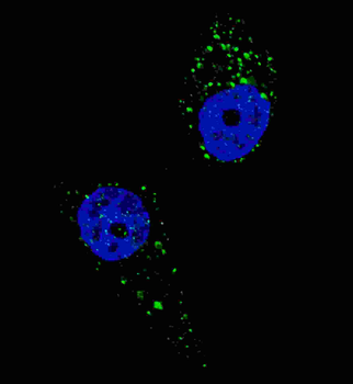
Fluorescent image of U251 cells stained with UVRAG (C-term) antibody. U251 cells were treated with Chloroquine (50 μM, 16 h), then fixed with 4% PFA (20 min), permeabilized with Triton X-100 (0.2%, 30 min). Cells were then incubated with UVRAG (C-term) primary antibody (1:200, 2 h at room temperature). For secondary antibody, Alexa Fluor 488 conjugated donkey anti-rabbit antibody (green) was used (1:1000, 1 h). Nuclei were counterstained with Hoechst 33342 (blue) (10 μg/ml, 5 min). UVRAG immunoreactivity is localized to autophagic vacuoles in the cytoplasm of U251 cells.
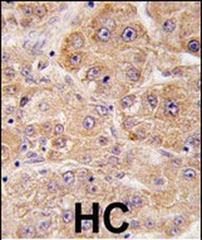
Formalin-fixed and paraffin-embedded human hepatocarcinoma tissue reacted with hUVRAG (C-term), which was peroxidase-conjugated to the secondary antibody, followed by DAB staining. This data demonstrates the use of this antibody for immunohistochemistry; clinical relevance has not been evaluated.
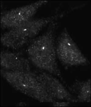
Immunofluorescence staining of Autophagy UVRAG antibody on Methanol-fixed HeLa cells.
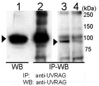
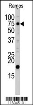
Immunoprecipitation and western blot analysis of anti-UVRAG Pab in 293T cells. UVRAG is immunoprecipitated (Lane 2) and detected in 293T cell transiently transfected with mouse UVRAG (Lane 1). Detection of endogenous UVRAG is shown in 293T cells (Lane 3) but is reduced by UVRAG siRNA transfection (Lane 4).
Quick Database Links
UniProt Details
− No UniProt data available
NCBI Reference Sequences
−Associated Accession Numbers
Curated reference sequences for the gene transcript and protein product| Protein | NP_003360.2 |
|---|
Documents Download
Datasheet
Product Information
Request a Document
Protocol Information
WB
Western Blot (IB, immunoblot)
IHC-P
Immunohistochemistry Paraffin
IF
Immunofluorescence
UVRAG Antibody (C-term) (orb1933088)

- 0.0
Based on 0 reviews
Participating in our Biorbyt product reviews program enables you to support fellow scientists by sharing your firsthand experience with our products.
Login to Submit a ReviewAvailable Sizes
Select a size below
Choose Conjugation or Carrier Free Version
Free Secondary Antibody (20 ul)0/0
Please add an antibody product to your cart first.