You have no items in your shopping cart.
Description
Research Area
Immunology & Inflammation
Images & Validation
−Item 1 of 7
| Tested Applications | FC |
|---|---|
| Reactivity | Human, Primate |
| Application Notes |
Key Properties
−| Antibody Type | Primary Antibody |
|---|---|
| Clonality | Monoclonal |
| Isotype | Mouse IgG1 kappa |
| Clone No. | 3G8 |
| Immunogen | Human neutrophils |
| Target | CD16 |
| Purification | Purified antibody is conjugated with fluorescein isothiocyanate (FITC) under optimum conditions and unconjugated antibody and free fluorochrome are removed by size-exclusion chromatography. |
| Conjugation | FITC |
Storage & Handling
−| Storage | Store at 2-8°C. Protect from prolonged exposure to light. Do not freeze. |
|---|---|
| Buffer/Preservatives | Stabilizing phosphate buffered saline (PBS), pH 7.4, 15 mM sodium azide |
| Expiration Date | 12 months from date of receipt. |
| Disclaimer | For research use only |
Alternative Names
−FcgammaRIII, IGFR3, FCRIII
Similar Products
−- Item 1 of 4
- Item 1 of 2
- Item 1 of 2


Quality Guarantee
Explore bioreagents carefree to elevate your research. All our products are rigorously tested for performance. If a product does not perform as described on its datasheet, our scientific support team will provide expert troubleshooting, a prompt replacement, or a refund. For full details, please see our Terms & Conditions and Buying Guide. Contact us at support@biorbyt.com.
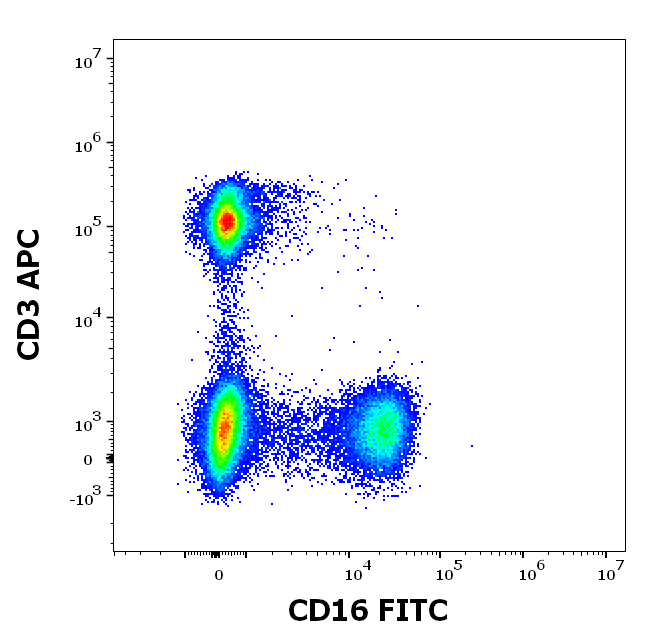
Staining pattern of Anti-human CD16 FITC antibody (clone 3G8) in dot-plot fluorescence visualization (lymphocyte gate) Analysis of the antibody staining profile was performed on blood leukocytes isolated from peripheral whole blood by bulk erythrocyte lysis using 10× diluted EXCELLYSE Live. Mouse monoclonal anti-human CD16 FITC antibody (clone 3G8) was used in concentration 9 µg/ml in stained stained blood sample (2 x 10^6 cells) and Mouse monoclonal anti-human CD3 APC antibody (clone UCHT1) in concentration 6 µg/ml, respectively.
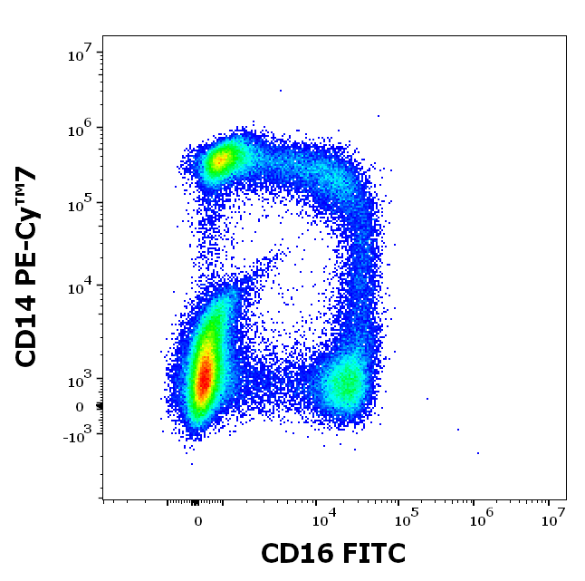
Staining pattern of Anti-human CD16 FITC antibody (clone 3G8) in dot-plot fluorescence visualization (mononuclear gate) Analysis of the antibody staining profile was performed on blood leukocytes isolated from peripheral whole blood by bulk erythrocyte lysis using 10× diluted EXCELLYSE Live. Mouse monoclonal anti-human CD16 FITC antibody (clone 3G8) was used in concentration 9 µg/ml in stained stained blood sample (2 x 10^6 cells) and Mouse monoclonal anti-human CD14 PE-Cy™7 antibody (clone MEM-15) in concentration 8 µg/ml, respectively.
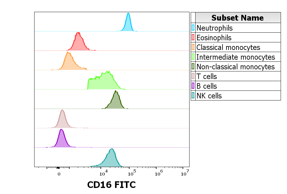
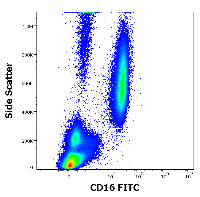
Reactivity of Anti-human CD16 FITC antibody (clone 3G8) on human peripheral leukocytes Analysis of the antibody staining profile was performed on blood leukocytes isolated from peripheral whole blood by bulk erythrocyte lysis using 10× diluted EXCELLYSE Live. Suspension of blood leukocytes (2 x 10^6 cells) was added to the mixture of CD16 FITC antibody (clone 3G8, 9 µg/ml in stained blood sample), backbone antibody conjugates and Monocyte Blocking Buffer, vortexed and incubated for 20 min. Stained sample was fixed with 2 ml of 10× diluted EXCELLYSE Easy solution for 10 min. Finally, samples were centrifuged (670 g, 5 min.), supernatant removed and the cell pellet was resuspended in 200 µl of PBS for acquisition.
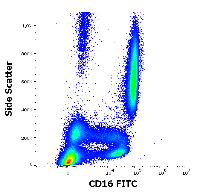
Anti-human CD16 FITC antibody (clone 3G8) works in flow cytometry application. Analysis of the antibody staining profile was performed on blood leukocytes isolated from peripheral whole blood by bulk erythrocyte lysis using 10× diluted EXCELLYSE Live. Mouse monoclonal anti-human CD16 FITC antibody (clone 3G8) was used in concentration 9 µg/ml in stained blood sample (2 x 10^6 cells).
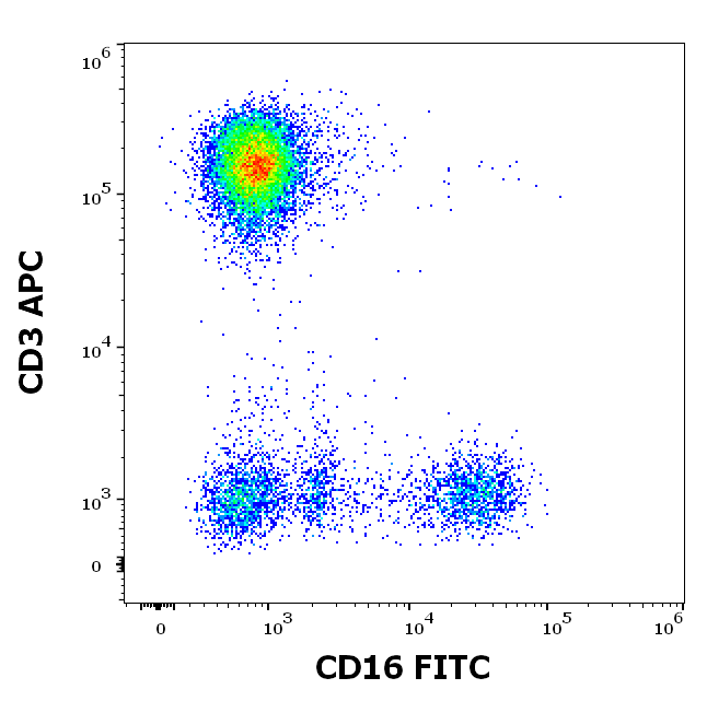
Flow cytometry multicolor surface staining pattern of human peripheral whole blood stained using anti-human CD16 (3G8) FITC antibody (4 μl reagent / 100 μl of peripheral whole blood) and anti-human CD3 (UCHT1) APC antibody (10 μl reagent / 100 μl of peripheral whole blood).
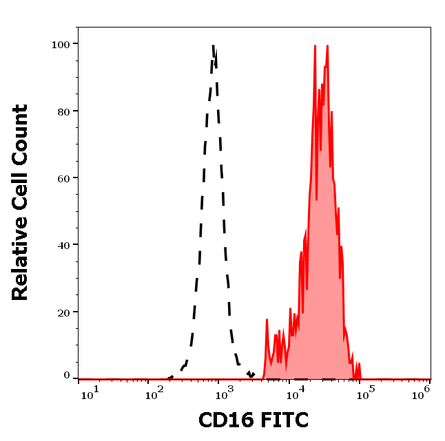
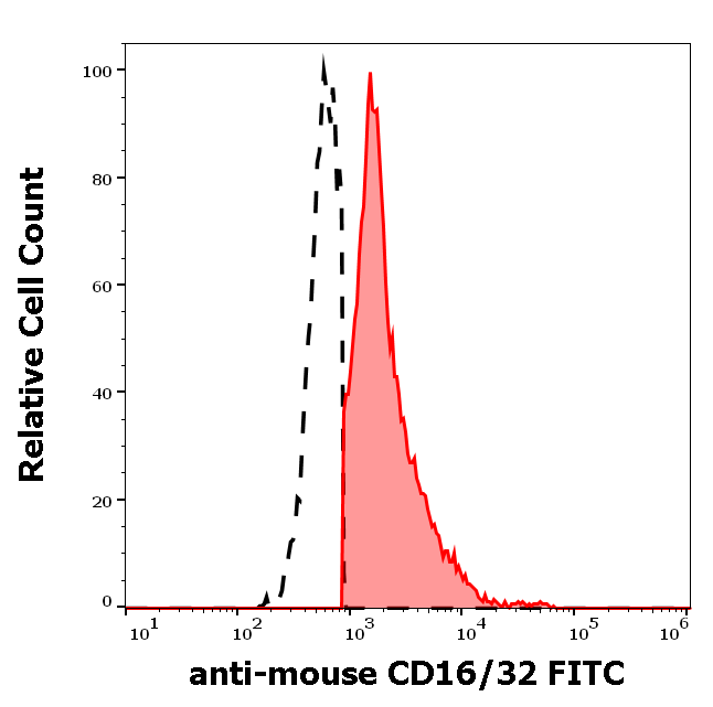
Separation of human CD16 positive CD3 negative lymphocytes (red-filled) from CD16 negative CD3 positive lymphocytes (black-dashed) in flow cytometry analysis (surface staining) of human peripheral whole blood stained using anti-human CD16 (3G8) FITC antibody (4 μl reagent / 100 μl of peripheral whole blood).
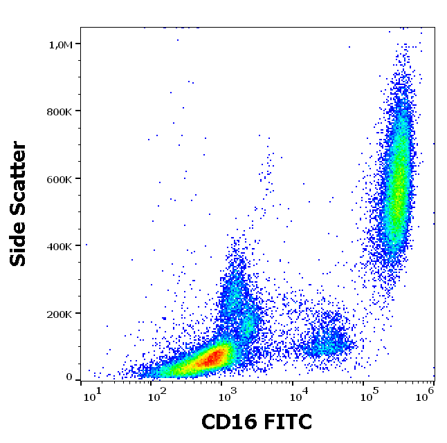
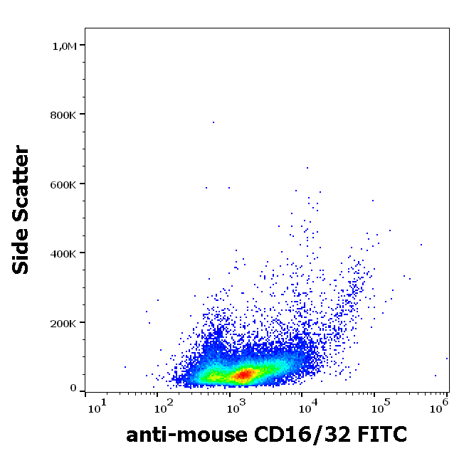
Flow cytometry surface staining pattern of human peripheral whole blood stained using anti-human CD16 (3G8) FITC antibody (4 μl reagent / 100 μl of peripheral whole blood).
Documents Download
Datasheet
Product Information
Request a Document
CD16 Antibody (FITC) (orb44655)

- 0.0
Based on 0 reviews
Participating in our Biorbyt product reviews program enables you to support fellow scientists by sharing your firsthand experience with our products.
Login to Submit a ReviewAvailable Sizes
Select a size below
Free Secondary Antibody (20 ul)0/0
Please add an antibody product to your cart first.