You have no items in your shopping cart.
Description
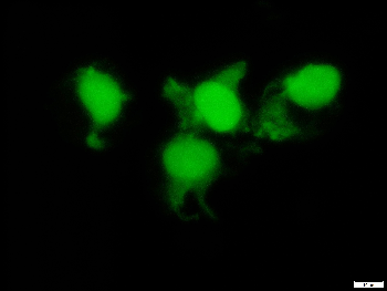
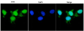
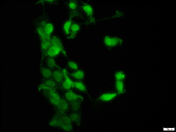
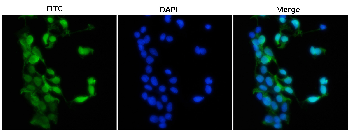
Images & Validation
−Item 1 of 3
| Tested Applications | FC, ICC, WB |
|---|---|
| Application Notes |
Key Properties
−| Antibody Type | Primary Antibody |
|---|---|
| Clonality | Polyclonal |
| Isotype | Rabbit polyclonal |
| Immunogen | mCherry protein from Anaplasma marginale |
| Target | mCherry |
| Purification | Purified by protein-A affinity chromatography. |
| Conjugation | Unconjugated |
Storage & Handling
−| Storage | Maintain refrigerated at 2-8°C for up to 2 weeks. For long term storage store at -20°C in small aliquots to prevent freeze-thaw cycles. |
|---|---|
| Buffer/Preservatives | Phosphate buffered saline (PBS), pH 7.4, 15 mM sodium azide |
| Concentration | 1 mg/ml |
| Expiration Date | 12 months from date of receipt. |
| Disclaimer | For research use only |
Similar Products
−- Item 1 of 2
tdTomato Antibody [orb182397]
ELISA, FACS, IF, IHC-Fr, IHC-P, WB
Other
Goat
Polyclonal
Unconjugated
100 μg - Item 1 of 4
mCherry Goat Polyclonal Antibody [orb11618]
FC, ICC, IEM, IF, IHC-Fr, IHC-P, WB
Other
Goat
Polyclonal
Unconjugated
20 μg, 100 μg - Item 1 of 6
mCherry Mouse Monoclonal Antibody [orb66657]
ELISA, FC, ICC, IF, WB
Other
Mouse
Monoclonal
Unconjugated
100 μg, 20 μg - Item 1 of 8
mCherry Chicken Polyclonal Antibody [orb611194]
ELISA, FC, ICC, IF, WB
Other
Gallus
Polyclonal
Unconjugated
200 μg, 100 μg, 20 μg - Item 1 of 6
mCherry Rabbit Polyclonal Antibody [orb181783]
ELISA, FC, ICC, IF, WB
Other
Rabbit
Polyclonal
Unconjugated
100 μg, 20 μg


Quality Guarantee
Explore bioreagents carefree to elevate your research. All our products are rigorously tested for performance. If a product does not perform as described on its datasheet, our scientific support team will provide expert troubleshooting, a prompt replacement, or a refund. For full details, please see our Terms & Conditions and Buying Guide. Contact us at support@biorbyt.com.
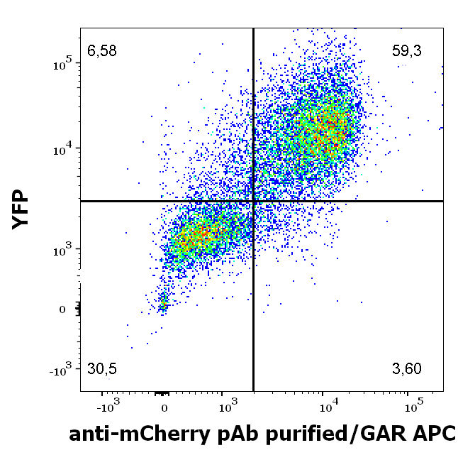
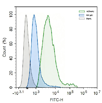
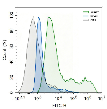
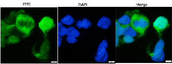
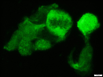
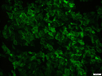
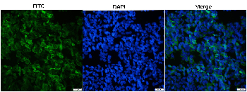
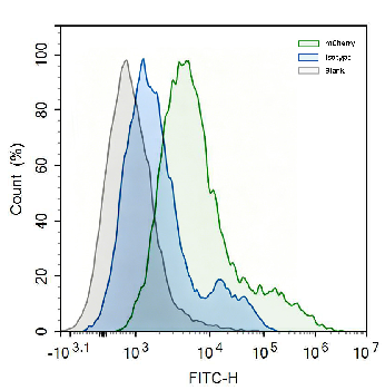
Flow cytometry surface staining pattern of HEK293T/17 cells co-transfected with mCherry/GPI and YFP/GPI constructs stained using anti-mCherry Purified rabbit polyclonal antibody (concentration in sample 2 μg/ml, GAR APC).
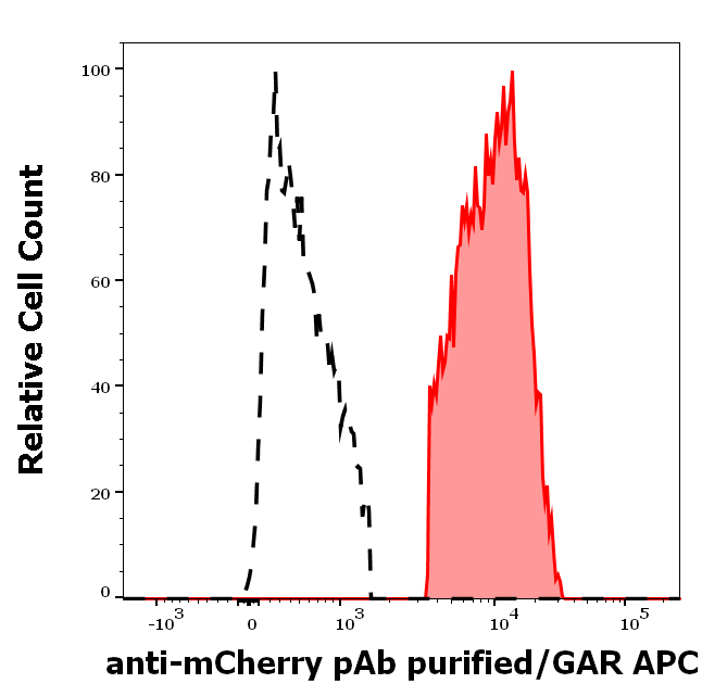
Separation of HEK293T/17 cells co-transfected with mCherry/GPI and YFP/GPI constructs stained anti-mCherry Purified rabbit polyclonal antibody (concentration in sample 2 μg/ml, GAR APC, red-filled) from HEK293T/17 cells co-transfected with mCherry/GPI and YFP/GPI constructs unstained by primary polyclonal antibody (GAR APC, black-dashed) in flow cytometry analysis (surface staining) of HEK293T17/mCherry/YFP cell suspension.

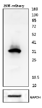
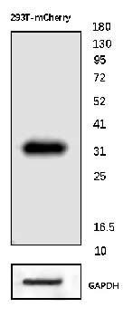
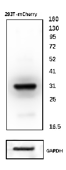
Western blotting analysis of mCherry fluorescent protein using rabbit polyclonal PAb (918) on lysates of HEK293T/17 cells co-transfected with mCherry/GPI and YFP/GPI constructs (HEK293T/17 cells transfected with YFP/GPI; negative control) under reducing and non-reducing conditions. Nitrocellulose membrane was probed with 2 µg/ml of rabbit anti-mCherry polyclonal antibody followed by IRDye800-conjugated anti-rabbit secondary antibody. A specific band was detected for mCherry protein at approximately 30 kDa.
Documents Download
Datasheet
Product Information
Request a Document
Protocol Information
WB
Western Blot (IB, immunoblot)
FC
Flow Cytometry
ICC
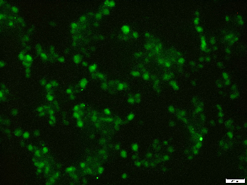
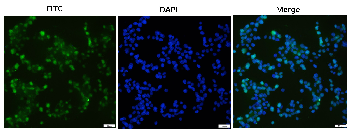
Immunocytochemistry
mCherry Antibody (orb763362)

- 0.0
Based on 0 reviews
Participating in our Biorbyt product reviews program enables you to support fellow scientists by sharing your firsthand experience with our products.
Login to Submit a ReviewAvailable Sizes
Select a size below
Free Secondary Antibody (20 ul)0/0
Please add an antibody product to your cart first.