You have no items in your shopping cart.
ATP6V1B1 Antibody
SKU: orb1270112
Featured
Description
Research Area
Metabolism Research, Signal Transduction
Images & Validation
−Item 1 of 3
| Tested Applications | IF, IHC-P, WB |
|---|---|
| Reactivity | Human |
| Predicted Reactivity | Bovine, C. elegans, Drosophila, Gallus, Mouse, Rat |
| Application Notes |
Key Properties
−| Antibody Type | Primary Antibody |
|---|---|
| Host | Rabbit |
| Clonality | Polyclonal |
| Isotype | Rabbit Ig |
| Immunogen | This ATP6V1B1 antibody is generated from rabbits immunized with a KLH conjugated synthetic peptide between 284-310 amino acids from the Central region of human ATP6V1B1. |
| Target | ATP6V1B1 |
| Molecular Weight | 57 kDa |
| Purification | This antibody is purified through a protein A column, followed by peptide affinity purification. |
| Conjugation | Unconjugated |
Storage & Handling
−| Storage | Maintain refrigerated at 2-8°C for up to 2 weeks. For long term storage store at -20°C in small aliquots to prevent freeze-thaw cycles. |
|---|---|
| Form/Appearance | Liquid |
| Buffer/Preservatives | Supplied in PBS with 0.09% (W/V) sodium azide. |
| Concentration | batch dependent |
| Expiration Date | 12 months from date of receipt. |
| Disclaimer | For research use only |
Alternative Names
−V-type proton ATPase subunit B, kidney isoform, V-ATPase subunit B 1, Endomembrane proton pump 58 kDa subunit, Vacuolar proton pump subunit B 1, ATP6V1B1, ATP6B1, VATB, VPP3
Similar Products
−- Item 1 of 3
ATP6V1B1 Antibody (Center) [orb1938126]
IF, IHC-P, WB
Bovine, C. elegans, Drosophila, Gallus, Mouse, Rat
Human
Rabbit
Polyclonal
Unconjugated
50 μl, 100 μl - Item 1 of 2
- Item 1 of 1
- Item 1 of 1
- Item 1 of 2

Quality Guarantee
Explore bioreagents carefree to elevate your research. All our products are rigorously tested for performance. If a product does not perform as described on its datasheet, our scientific support team will provide expert troubleshooting, a prompt replacement, or a refund. For full details, please see our Terms & Conditions and Buying Guide. Contact us at support@biorbyt.com.

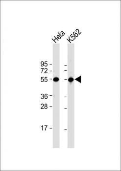
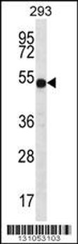
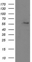
Western Blot at 1:1000 dilution Lane 1: Hela whole cell lysate Lane 2: K562 whole cell lysate Lysates/proteins at 20 ug per lane.
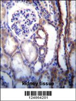
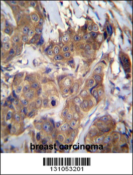
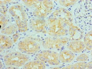
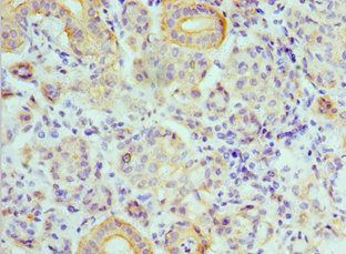
ATP6V1B1 Antibody immunohistochemistry analysis in formalin fixed and paraffin embedded human kidney tissue followed by peroxidase conjugation of the secondary antibody and DAB staining.

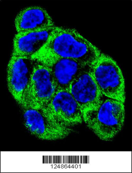
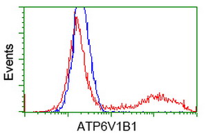
Confocal immunofluorescent analysis of ATP6V1B1 Antibody with WiDr cell followed by Alexa Fluor 488-conjugated goat anti-rabbit lgG (green). DAPI was used to stain the cell nuclear (blue).
Documents Download
Datasheet
Product Information
Request a Document
Protocol Information
WB
Western Blot (IB, immunoblot)
IHC-P
Immunohistochemistry Paraffin
IF
Immunofluorescence
ATP6V1B1 Antibody (orb1270112)

- 0.0
Based on 0 reviews
Participating in our Biorbyt product reviews program enables you to support fellow scientists by sharing your firsthand experience with our products.
Login to Submit a ReviewAvailable Sizes
Select a size below