You have no items in your shopping cart.
Description
Images & Validation
−Item 1 of 4
| Tested Applications | ELISA, IHC, WB |
|---|---|
| Dilution Range | ELISA: 1:20,000, IHC: 1.25-2.5 µg/ml, WB: 1:1,000 - 1:5,000 |
| Reactivity | Human, Mouse |
| Application Notes |
Key Properties
−| Antibody Type | Primary Antibody |
|---|---|
| Host | Rabbit |
| Clonality | Polyclonal |
| Isotype | IgG |
| Immunogen | This affinity purified antibody was prepared from whole rabbit serum produced by repeated immunizations with a synthetic peptide corresponding to a region near the amino terminus of mouse Sipa1. |
| Target | Sipa1 |
| Purity | This product was affinity purified from monospecific antiserum by immunoaffinity chromatography. This antibody is specific for mouse Sipa1 protein. A BLAST analysis was used to suggest cross-reactivity with Sipa1 from mouse, human and rat based on a 100% homology with the immunizing sequence. Cross-reactivity with Sipa1 from other sources has not been determined. |
| Conjugation | Unconjugated |
Storage & Handling
−| Storage | Store vial at -20° C prior to opening. Aliquot contents and freeze at -20° C or below for extended storage. Avoid cycles of freezing and thawing. Centrifuge product if not completely clear after standing at room temperature. This product is stable for several weeks at 4° C as an undiluted liquid. Dilute only prior to immediate use. |
|---|---|
| Form/Appearance | Liquid (sterile filtered) |
| Buffer/Preservatives | Preservative: 0.01% (w/v) Sodium Azide. Stabilizer: None; Buffer: 0.02 M Potassium Phosphate, 0.15 M Sodium Chloride, pH 7.2 |
| Concentration | 1.25 mg/mL |
| Expiration Date | 12 months from date of receipt. |
| Dry Ice Shipping | Please note: This product requires shipment on dry ice. A dry ice surcharge will apply. |
| Disclaimer | For research use only |
Alternative Names
−rabbit anti-Sipa1 antibody, Sipa-1, Sipa 1, GTPase activating protein Spa 1 antibody, p130 SPA-1 antibody, Signal induced proliferation associated 1 antibody, SPA1
Similar Products
−- Item 1 of 2
SIPA1L3 Antibody [orb1538477]
ELISA, IF, IHC, IHC-P, WB
Human, Mouse, Rat
Rabbit
Polyclonal
Unconjugated
0.05 mg - Item 1 of 2
- Item 1 of 2
- Item 1 of 2
- Item 1 of 3
SIPA1L3 Antibody [orb1238968]
ELISA, IF, IHC-P, WB
Human, Mouse, Rat
Rabbit
Polyclonal
Unconjugated
0.1 mg, 0.02 mg


Quality Guarantee
Explore bioreagents carefree to elevate your research. All our products are rigorously tested for performance. If a product does not perform as described on its datasheet, our scientific support team will provide expert troubleshooting, a prompt replacement, or a refund. For full details, please see our Terms & Conditions and Buying Guide. Contact us at support@biorbyt.com.
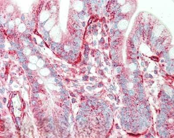
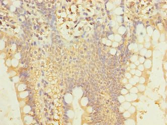
Immunohistochemistry of rabbit anti-Sipa1 antibody. Tissue: small intestine. Fixation: formalin fixed paraffin embedded. Antigen retrieval: not required. Primary antibody: Anti-Sipa1 at 5 µg/ml for 1 h at RT. Secondary antibody: Peroxidase rabbit secondary antibody at 1:10000 for 45 min at RT. Staining: Sipa-1 as precipitated red signal with hematoxylin purple nuclear counterstain.
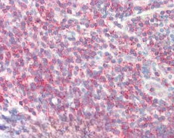
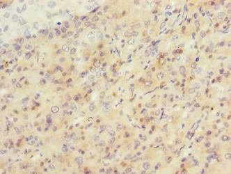
Immunohistochemistry of rabbit anti-Sipa1 antibody. Tissue: tonsil. Fixation: formalin fixed paraffin embedded. Antigen retrieval: not required. Primary antibody: Anti-Sipa1 at 5 µg/ml for 1 h at RT. Secondary antibody: Peroxidase rabbit secondary antibody at 1:10000 for 45 min at RT. Staining: Sipa-1 as precipitated red signal with hematoxylin purple nuclear counterstain.
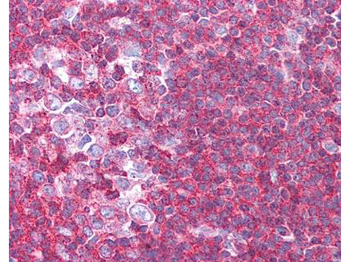
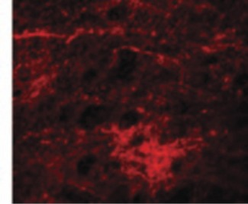
Biorbyt's affinity purified anti-Sipa1 antibody was used at 1.25 ug/ml to detect signal in a variety of tissues including multi-human, multi-brain and multi-cancer slides. This image shows moderate to strong positive staining of lymphocytes within human tonsil at 40X. Tissue was formalin-fixed and paraffin embedded. The image shows localization of the antibody as the precipitated red signal, with a hematoxylin purple nuclear counterstain.
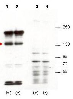
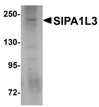
Western blot using Biorbyt's affinity purified anti-Sipa1 antibody shows detection of over-expressed Sipa1 in lysates from mouse 3T3 cells transfected with Sipa1 (lane 1). Endogenous Sipa1 is detected in lane 2, which contains lysate from 3T3 cells mock-transfected with LacZGLB, although at a significantly reduced level compared to transfected cells. Lane 3 and 4 are similar to lanes 1 and 2 except the antibody was preincubated with the immunizing peptide prior to reaction with the membrane. The identity of the higher and lower molecular weight bands is unknown. The band at ~130 kDa, indicated by the arrowhead, corresponds to recombinant Sipa1. Primary antibody was used at 1:1250.
Quick Database Links
UniProt Details
− No UniProt data available
NCBI Reference Sequences
−Associated Accession Numbers
Curated reference sequences for the gene transcript and protein product| RefSeq | AAH54824.1 |
|---|
Documents Download
Datasheet
Product Information
Request a Document
Protocol Information
WB
Western Blot (IB, immunoblot)
IHC
Immunohistochemistry
ELISA
Enzyme-linked Immunosorbent Assay (EIA)
Sipa1 Antibody (orb345595)

- 0.0
Based on 0 reviews
Participating in our Biorbyt product reviews program enables you to support fellow scientists by sharing your firsthand experience with our products.
Login to Submit a ReviewAvailable Sizes
Select a size below
Choose Conjugation or Carrier Free Version
Free Secondary Antibody (20 ul)0/0
Please add an antibody product to your cart first.