You have no items in your shopping cart.
Description
Research Area
Immunology & Inflammation, Stem Cell & Developmental Biology
Images & Validation
−

Item 1 of 7
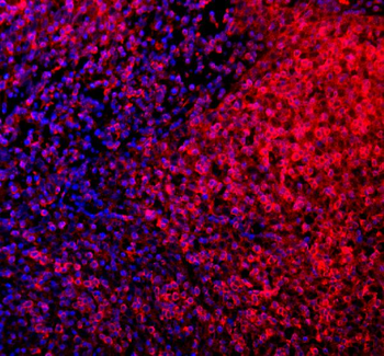
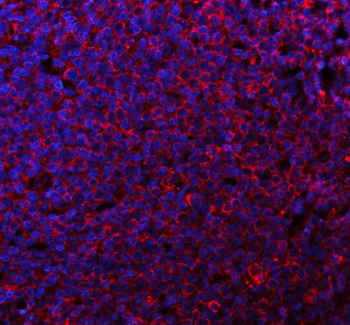
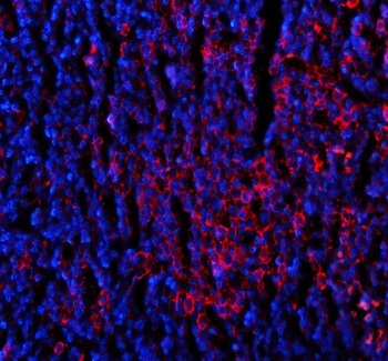
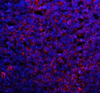
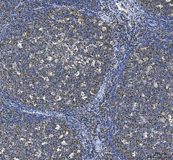
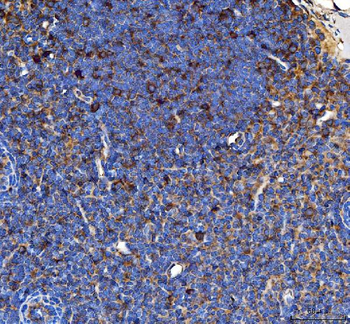
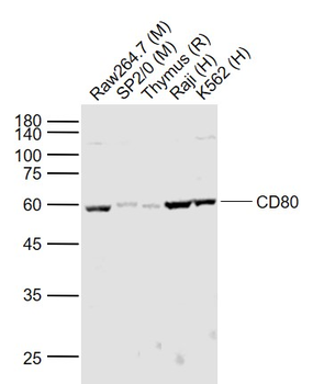
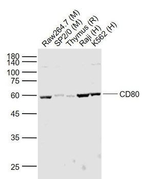
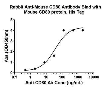
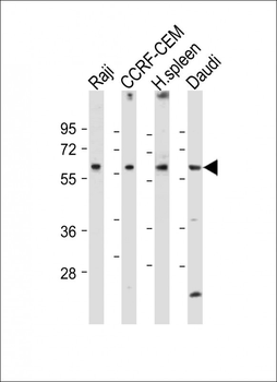
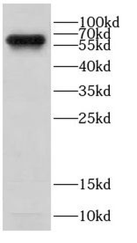
| Tested Applications | ELISA, IHC, WB |
|---|---|
| Dilution Range | Western blot, 0.1-0.5 μg/ml, Human, Mouse Immunohistochemistry (Paraffin-embedded Section), 2-5μg/ml, Human, Mouse, Rat ELISA, 0.1-0.5μg/ml |
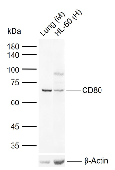
| Reactivity | Human, Mouse, Rat |
Related Conjugates & Formulations
−Key Properties
−| Antibody Type | Primary Antibody |
|---|---|
| Host | Rabbit |
| Clonality | Polyclonal |
| Isotype | Rabbit IgG |
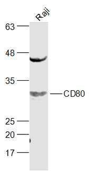
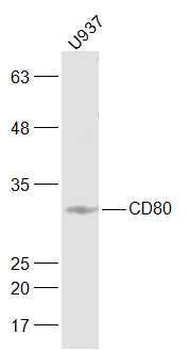
| Immunogen | E. coli-derived human CD80 recombinant protein (Position: V35-L244). |
| Target | T-lymphocyte activation antigen CD80 |
| Molecular Weight | 33-70 kDa |
| Purification | Immunogen affinity purified. |
| Conjugation | Unconjugated |
Storage & Handling
−| Storage | Maintain refrigerated at 2-8°C for up to 2 weeks. For long term storage store at -20°C in small aliquots to prevent freeze-thaw cycles. |
|---|---|
| Form/Appearance | Lyophilized |
| Buffer/Preservatives | Each vial contains 4 mg Trehalose, 0.9 mg NaCl and 0.2 mg Na2HPO4. |
| Concentration | Adding 0.2 ml of distilled water will yield a concentration of 500 μg/ml. |
| Expiration Date | 12 months from date of receipt. |
| Disclaimer | For research use only |
Alternative Names
−Activation B7 1 antigen; B7; B7 1; B7-1; BB1; CD28LG; CD28LG1; CD80; CD80 molecule; CTLA 4 counter receptor B7.1; LAB7
Similar Products
−- Item 1 of 2
CD80 Rabbit Polyclonal Antibody [orb5805]
FC, WB
Rat
Human, Mouse, Rat
Rabbit
Polyclonal
Unconjugated
50 μl, 100 μl, 200 μl - Item 1 of 3
CD80 Rabbit Polyclonal Antibody [orb500681]
IF, IHC-Fr, IHC-P, WB
Human
Rabbit
Polyclonal
Unconjugated
50 μl, 100 μl, 200 μl - Item 1 of 3
CD80 Rabbit Polyclonal Antibody [orb1675375]
ELISA, WB
Rat
Human, Mouse
Rabbit
Polyclonal
Unconjugated
50 μl, 100 μl, 200 μl - Item 1 of 2
- Item 1 of 2
CD80 Rabbit Polyclonal Antibody [orb625625]
ELISA, FC, IF, WB
Human, Mouse
Rabbit
Polyclonal
Unconjugated
50 μg, 100 μg

Quality Guarantee
Explore bioreagents carefree to elevate your research. All our products are rigorously tested for performance. If a product does not perform as described on its datasheet, our scientific support team will provide expert troubleshooting, a prompt replacement, or a refund. For full details, please see our Terms & Conditions and Buying Guide. Contact us at support@biorbyt.com.
Quick Database Links
Gene Symbol
T-lymphocyte activation antigen CD80
UniProt
UniProt Details
− No UniProt data available
Protocol Information
WB
Western Blot (IB, immunoblot)
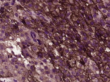
IHC
Immunohistochemistry
ELISA
Enzyme-linked Immunosorbent Assay (EIA)
Filter by Applications
Filter by Species
Wei-Shan Hsieh, Chia-Chi Kung, Shir-Ly Huang, Shih-Chang Lin, Wei-Hsin Sun TDAG8, TRPV1, and ASIC3 involved in establishing hyperalgesic priming in experimental rheumatoid arthritis Scientific Reports, 7, 8870 (2017)
Applications
IHC
Reactivity
Mouse
Available Sizes
Select a size below
Free Secondary Antibody (20 ul)0/0
Please add an antibody product to your cart first.